Loading...

Archive

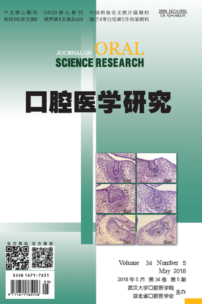
    28 May 2018, Volume 34 Issue 5 Previous Issue    Next Issue

    For Selected: Toggle Thumbnails
    Endoplasmic Reticulum Stress and Autophagy in the Study of Oral Diseases.
    LIU Jian-guo, JING Pei
    2018, 34(5): 461-466.  DOI: 10.13701/j.cnki.kqyxyj.2018.05.001
    Abstract ( 365 )   PDF (891KB) ( 357 )  
    Endoplasmic reticulum (ER) is a multifauncional oranelle responsible for intracellular protein synthesis and calcium storage, involved in various physiological activities. Mutiple external stimuli such as hypoxia stress, toxic substances and so on will lead to the endoplasmic reticulum dysfunction, resulting in unfolded and/or misfolded proteins, calcium homeostasis imbalance, cells damage and physiological function disorder, which will trigger many relative diseases. Autophagy is a cytoprotective mechanism, removing intracellular damaged organelles and unfolded/misfolded proteins, which can relief endoplasmic reticulum stress, restore cell homeostasis and maintain or improve cell viability by providing new marerials and energy. Recent sudies have shown that the interaction between endoplasmic reticulum stress and autophagy may plays a critical role in the development of multiple diseases. This review summarizes the roles of endoplasmic reticulum stress and autophagy in oral diseases.
    References | Related Articles | Metrics
    Research Progress in Photodynamic Therapy for Peri-implantitis.
    YANG Hai-li, ZOU Hai-xiao, SONG Li
    2018, 34(5): 467-469.  DOI: 10.13701/j.cnki.kqyxyj.2018.05.002
    Abstract ( 372 )   PDF (796KB) ( 576 )  
    Peri-implantitis is one of the most common complications after dental implantation, which results in the loosing and falling of the implant. It is a complex disease caused by a variety of anaerobic bacteria, and the treatment includes non-surgical and surgical methods. However, both of them have some limitations. Since 1990s, photodynamic therapy (PDT) has gained increasing attention as an alternative treatment approach. This review summarizes the mechanism and influence factor of PDT in the treatment of peri-implantitis, which may provide a guideline for the clinicians.
    References | Related Articles | Metrics
    Research Progress of Glass Ceramics Endocrowns Reconstructing Endodontically Treated Posterior Teeth.
    LU Jun-qing, ZHANG Xiu-yin
    2018, 34(5): 470-473.  DOI: 10.13701/j.cnki.kqyxyj.2018.05.003
    Abstract ( 308 )   PDF (809KB) ( 533 )  
    Restoration of endodontically treated posterior teeth with extensive coronal loss is still a clinical problem. With the development of dentistry material and widely employment of computer-aided design/manufacturing (CAD/CAM), endocrowns combined with high-strength glass ceramics, CAD/CAM, and reliable adhesive systems are widely accepted by both patients and prosthodontists for their minimal invasion and esthetic characteristics. This paper reviews the research progress on the comparison between endocrowns and conventional restoration, marginal preparation design, adaptation of endocrowns,ceramic materials,application of FRCs, and prognostic evaluation.
    References | Related Articles | Metrics
    Influence of Physicochemical Property of Hydrogel on Stem Cell Behavior.
    YANG Xue, GAO Hua-li, ZHANG Lu-lu, JIANG Qiu, Jin Zhi-en, WANG Ting-ting, XU Gao-xiang
    2018, 34(5): 474-476.  DOI: 10.13701/j.cnki.kqyxyj.2018.05.004
    Abstract ( 273 )   PDF (794KB) ( 408 )  
    Hydrogel is a kind of high molecular polymer which has a variety of hydrophilic groups and forms three-dimensional network structure. Hydrogel can construct extracellular microenvironment, and provide extracellular matrix for the adhesion, proliferation, differentiation and migration of stem cells. However, the behavior of the stem cells in hydrogel is influenced by many factors. Past researches mainly focused on the effect of biochemical signals on stems cells, however, more and more researchers begin to pay attention to the physicochemical properties of the hydrogel itself. In this paper, the effect of physicochemical properties of hydrogels on the behavior of stem cell was discussed.
    References | Related Articles | Metrics
    Research Progress of Gingival Papilla Reconstruction.
    ZHU Juan-juan, FENG Yun-xia
    2018, 34(5): 477-479.  DOI: 10.13701/j.cnki.kqyxyj.2018.05.005
    Abstract ( 387 )   PDF (788KB) ( 475 )  
    Loss of gingival papilla will lead to the formation of black triangle, which is not only an important factor affecting the aesthetics of oral soft tissue, but also affects the pronunciation and function. The patients are urgent to solve the problem. The methods to reconstruct the gingival papilla include surgery and non surgery ways. Non surgery ways include orthodontic treatment, prosthetic treatment, and injection of hyaluronic acid. This article gives a summary on the research progress of gingival papilla reconstruction.
    References | Related Articles | Metrics
    Phenotypic Observation of Mandibular First Molar in Caveolin-1 Gene Knockout Mice.
    SHI Lu, TIAN Fei-fei, BAI Shu-feng, FENG Hai-lin
    2018, 34(5): 480-484.  DOI: 10.13701/j.cnki.kqyxyj.2018.05.006
    Abstract ( 202 )   PDF (2966KB) ( 258 )  
    Objective: To investigate the histological morphology of mandibular first molar tooth germ and analyse dental phenotype in caveolin-1 gene knockout (KO) mice. Methods: After genotype identification with polymerase chain reaction (PCR), mandibular first molar germs and teeth were collected from caveolin-1 gene knockout mice and wild-type (WT) homologous mice. The histological morphology of tooth germs and dental phenotype were studied by hematein eosin(HE)staining and micro computed tomography (Micro-CT),respectively. Results:At E14.5, the tooth germ of WT mouse was at the cap stage; however, the tooth germ of KO mouse was still at the bud stage, and no obvious stratification and primary enamel knot were found. With the tooth germ development, the enamel organ cells of KO mouse stratified into four layers at cap stage (E16.5). While, enamel organ of KO mouse had smaller volume and less invagination of cervical loop into epithelial mesenchymal. At E18.5, the cell polarities of both ameloblasts and odontoblasts were not obvious in KO tooth germ. And the odontoblasts were lack of typical high columnar shapes. The results of Micro-CT and 3D reconstruction showed that no significant difference was found between the two mice’s lower first molar anatomic shape. Conclusion: The deficiency of caveolin-1 gene led to temporal arrest for the bud-to-cap transition in developmental molar tooth germs without significant influence on the later developmental processes.
    References | Related Articles | Metrics
    Analysis of Metabonomics on Maternal Plasma in Mice with Cleft Palate Induced by Dexamethasone.
    WANG Shi-lin, YU Zi-jing, HE Wei
    2018, 34(5): 485-489.  DOI: 10.13701/j.cnki.kqyxyj.2018.05.007
    Abstract ( 218 )   PDF (1267KB) ( 189 )  
    Objective: To discover the difference of dexamethasone group and control group of pregnant mice in plasma metabolites during the embryonic palate development, and to lay foundations for discovering the specific metabolites during dexamethasone-induced cleft palate. Methods: Pregnant mice were collected and randomly divided into dexamethasone group (experimental group) and control group. During the specific pregnancy, the mice were peritoneally injected with certain amount of dexamethasone and saline respectively. After pregnant mice were sacrificed, plasma samples were collected in specific pregnancy and the incidence of mice cleft palate was estimated. Nuclear magnetic resonance spectrometer was applied to get the nuclear magnetic resonance hydrogen spectrum diagram of pregnant mice plasma samples. When combined with principal component analysis and partial least squares depict the metabolic differences between the experimental group and the control group of pregnant mice plasma metabolites in the development stage of embryonic palate, specific metabolic markers of cleft palate induced by dexamethasone were obtained. Results: The experimental group and the control group were different in plasma metabolites during embryonic palatal development. Conclusion: Dexamethasone may cause changes in amino acid signal during the embryonic palatal fusion phase, and activate the related regulation mechanism, which delayed the embryonic development and resulted in cleft palate.
    References | Related Articles | Metrics
    Construction of Streptococcus Mutans Fluoride-resistant Strain with GtfB Gene Inactive.
    LI Wen-yue, GAO Shuang, ZHANG Zhi-ying, ZHANG Hong, CHAO Bo, WU Yu-feng, LI He, WANG Chun-meng, ZHANG Zhi-min
    2018, 34(5): 490-494.  DOI: 10.13701/j.cnki.kqyxyj.2018.05.008
    Abstract ( 215 )   PDF (1285KB) ( 199 )  
    Objective: To construct Streptococcus mutans (S.mutans) fluoride-resistant strain UA159-FR with gtfB gene inactive to investigate the function of gtfB gene in fluoride-resistant strain. Methods: The S.mutans fluoride-resistant strain UA159-FR was cultured and used as a template to amplify the upstream and downstream homologous arm fragments of the gtfB gene. The kan gene was amplified by using the plasmid pEGFP-N1 as a template. Homologous recombination fragments of three fragments were obtained by overlap extension polymerase chain reaction (OE-PCR) and ligated with pEASY-Blunt Cloning Vector to form recombinant plasmids. The recombinant plasmids were identified by PCR and sequencing. The recombinant fragment was electrotransformed into UA159-FR competent cells to obtain the inactivated strain and identified by PCR. Results: The recombinant plasmids were successfully constructed via being identified by PCR and sequencing. It was identified by PCR that UA159-FR with gtfB gene inactive was obtained. Conclusion: The recombinant fragments of gtfB gene of S.mutans with its recombinant plasmids and the S.mutans fluoride-resistant strain UA159-FR with gtfB gene inactive were successfully constructed and could be used to study the function of gtfB gene.
    References | Related Articles | Metrics
    Effects of Porphyromonas Gingivalis Infection on Placental Tissue and Birth Weight of Neonates in Rats.
    LIANG Shan-shan, XING Wen-yan, JI Yao-ting, JIANG Han, DU Min-quan
    2018, 34(5): 495-499.  DOI: 10.13701/j.cnki.kqyxyj.2018.05.009
    Abstract ( 230 )   PDF (2252KB) ( 236 )  
    Objective: To investigate the effects of Porphyromonas gingivalis infection on expressions of placental Fas ligand (Fas L), Toll-like receptor (TLR)2 and TLR4, and birth weight of neonates in rats. Methods: Female rats were randomly divided into 3 groups: P.gingivalis-infected group, negative control group, and blank control group (n=13 for each group). Murine model of P.gingivalis infection was established by the tail vein injection. Birth weight of every neonate and litter weights was recorded, and the placentas were obtained at gestational day of 18. The expression levels of placental Fas, FasL, TLR2, and TLR4 were detected by Western blot analysis and immunohistochemical staining. Results: The newborn rats showed significantly lower birth weight in P.gingivalis-infected group. Furthermore, Fas, FasL, and TLR4 in placental tissues were significantly increased following P.gingivalis infection. However, the expression of TLR2 had no significant difference between P.gingivalis-infected group and negative control group. Conclusion: In our murine model, with elevated expression of Fas, FasL and TLR4 in placenal tissues, P.gingivalis infection induces lower birth weight of neonates. The results indicate that pregnant woman suffering from periodontal disease, with delivery of low birth weight babies, might be related to P.gingivalis-induced abnormally regulation of placental Fas, FasL and TLR4.
    References | Related Articles | Metrics
    Effect of p-Akt on Apoptosis of Human Extravillous Trophoblast Cells Induced by Porphyromonas Gingivalis.
    GUO Hai-ying, REN Hong-yu, JI Yao-ting, JIANG Han, DU Min-quan
    2018, 34(5): 500-504.  DOI: 10.13701/j.cnki.kqyxyj.2018.05.010
    Abstract ( 185 )   PDF (1672KB) ( 242 )  
    Objective: To study the effect of Porphyromonas gingivalis on human extravillous trophoblast cell line (HTR-8) and the role of p-Akt in this process. Methods: HTR-8 cells were infected with Porphyromonas gingivalis ATCC 33277 at a multiplicity of infection (MOI) of 200 or 0. The target siRNA was chosen to inactivate PI3K, inverted microscope and confocal microscope were used to record the morphological changes of HTR-8 cells, flow cytometry was used to detect the apoptosis of HTR-8 cells after infection with Porphyromonas gingivalis, and real time-PCR was used to test the inactivation of siRNA whose target was PI3K. Results: When HTR-8 cells were infected with Porphyromonas gingivalis for 12 hours, there was no statistical significance of the apoptosis rate in MOI 200 group compared with MOI 0 group. HTR-8 cells were infected for 24 hours, the number of apoptotic cells in MOI 200 group significantly increased (P<0.01). Infected for 48 hours, the extent of apoptosis was more obvious compared with control group MOI 0 group (P<0.001). When HTR-8 cells were pretreated with siRNA and then co-cultured with Porphyromonas gingivalis for 48 hours (SI 200 Group), the relative amounts of PI3K mRNA in SI 200 group was much lower than that in interference group (C 200 group) (P<0.01). At the same time, the apoptosis rate of SI 200 group was greater than 45%, which was significantly higher than that of MOI 200 group (P<0.01); and there was no statistical difference of apoptosis rate between MOI 200 group and C 200 group. Conclusion: Porphyromonas gingivalis does induce apoptosis in human extravillous trophoblast cells and p-Akt is activated to inhibit the apoptosis of HTR-8 cells to some extent in this process. All of these suggest that the mechanism by which periodontitis promotes preterm low birth weight is that Porphyromonas gingivalis induced human extravillous trophoblastic cells apoptosis.
    References | Related Articles | Metrics
    Polymerization Shrinkage and Mechanical Property of Light Curing Composite Resin Incorporated a Novel Monovinyl Acrylates Monomer.
    ZHANG Na, XIE Chao, HE Hui-ming, GONG Xu
    2018, 34(5): 505-508.  DOI: 10.13701/j.cnki.kqyxyj.2018.05.011
    Abstract ( 288 )   PDF (1140KB) ( 369 )  
    Objective: To evaluate the polymerization shrinkage and mechanical property of light curing composite resin incorporated new monovinyl acrylates monomer. Methods: New monovinyl acrylates monomer was incorporated into traditional composite resin as experimental group. The composite resin used TEGDMA was served as control group 1. Tentric N-Ceram, Neofil, and GC were selected as control group 2, 3, and 4, respectively. The X-ray micro-computed tomography was used to determine the volume of five composite resins after polymerization shrinkage. The mechanical properties of the samples were measured with universal testing machine and microhardness meter. The polymerization shrinkage, flexural strength, compressive strength, diametral tensile strength, and microhardness were measured and compared. Results: The polymerization shrinkage of the light curing composite resin incorporated new monovinyl acrylates monomer was decreased in contrast to the control group 1. There were no significant differences in polymerization shrinkage between the experimental group and the control group 2-4. There were no significant differences in mechanical properties between the experimental group and the four control groups. Conclusion: The incorporation of monovinyl acrylates monomer possessed the potential to replace TEGDMA as a reactive diluent in dental resin which displayed favorable and unique properties. The material has its value in clinical application.
    References | Related Articles | Metrics
    Effects of Nano-particles on Propertites of Glass-ionomer Cement.
    SUN Jing, ZHU Bo-wu, YANG Lei, LIN Yong-sheng, CAO Bao-cheng
    2018, 34(5): 509-512.  DOI: 10.13701/j.cnki.kqyxyj.2018.05.012
    Abstract ( 197 )   PDF (2127KB) ( 279 )  
    Objective: To explore the effect of co-doping with TiO2 nanoparticles and sisal cellulose nanocrystals (CNCs) on the physical and antibacterial properties of a conventional glass-ionomer cement (GIC). Methods: Test samples were divided into three groups: 2wt.% titanium dioxide group (T), 2wt.% titanium dioxide + 0.4wt.% CNCs co-doped group (C), and unmodified cement control group (CG). The samples were subjected to mechanical tests to evaluate the compressive strength and wear resistance. Antimicrobial activity was evaluated against candida albicans. Results: Compared with the control group, the co-doped group demonstrated an increased compressive strength of 21.8%(P<0.001), the volume wear rate was reduced to 35.9% (P<0.001). The antibacterial effect against candida albicans was increased to 92.3%(P<0.001). Scanning electron microscopy showed a significant reduction of cracks in group C. Conclusion: The co-doping of CNCs and TiO2 nanoparticles significantly improve the compressive properties, abrasion resistance, and antibacterial effect of GIC.
    References | Related Articles | Metrics
    Biomechanical Analysis of Repairing Large Area Defects of Mandibular First Molar with Endocrown and Post-core Crown.
    ZHAO Chu-qiao, XU Yi-chi, LIU Ding-kun, YANG Jun-xing, WEI Zi-qing, TANG Lin-jun, SHEN Jie, LIU Yu-kun, LIU Zhi-hui
    2018, 34(5): 513-517.  DOI: 10.13701/j.cnki.kqyxyj.2018.05.013
    Abstract ( 225 )   PDF (1144KB) ( 296 )  
    Objective: To compare the stress magnitude and distribution of residual dentin in mandibular first molar restored with endocrown or post-core crown using three-dimension finite element methods. Methods: The three-dimensional finite element models of mandibular first molar were established after scanning by CT. A complete tooth was set as the control group. After simulating root canal therapy, three experimental group models with three kinds of defects were set as the experimental group: defect of mesial and distal proximal surface (group A); defect of buccal and mesial proximal surface (group B); defect of buccal, mesial, and distal proximal surface (group C). Then, endocrowns and post-core crowns were used to restore the defects. A load of 200N, simulating intercuspal occlusion, was applied vertically to the occlusal surface, and a load of 200 N simulating lateral occlusion was applied to the occlusal surface with a 45°angle to the long axis of the tooth. Von Mises stresses and max principal stresses were calculated by Abaqus software. Results: The stress of tooth tissue restored by endocrown was higher than that of post-core crown even more than 1 times. The stress of endocrown was concentrated in the pulp floor, and that of the post-core crown was concentrated in the distal apical 1/3. Conclusion: Endocrown and post-core crown are both applicable for mandibular first molar with large area defect, however, the post-core crown has better protective effect for the tooth tissue.
    References | Related Articles | Metrics
    Marginal Fitness of Three Types of CAD/CAM Customized Abutments between Implant-abutment Interfaces.
    ZHU Qing, ZHU Chen-yuan, SHE Wen-jun, JIAO Ting
    2018, 34(5): 518-521.  DOI: 10.13701/j.cnki.kqyxyj.2018.05.014
    Abstract ( 245 )   PDF (2015KB) ( 255 )  
    Objective: To compare the marginal fitness of three types of CAD/CAM customized implant abutments (one-piece zirconia abutments, two-piece zirconia abutments and one-piece titanium abutments) between the implant-abutment interfaces. Methods: 3SHAPE software was used to design the customized CAD/CAM abutments. Three types of implants including one-piece zirconia abutments, two-piece zirconia abutments, and one-piece titanium abutment were fabricated after importing the designed data into computer-controlled cutting system (n=15). Stereomicroscope was used to measure the micro-gap of the implant-abutment interface. The results of the gap value were analyzed using statistical method. Results: The titanium abutment showed the lowest gap values, and those of the two-piece zirconia abutments were lower than those of the one-piece zirconia abutments. There was no statistical difference on the gap values among the palatal, labial, mesial, and distal surfaces in the same type of abutment. Conclusion: The one-piece titanium abutment showed the best marginal fit.
    References | Related Articles | Metrics
    Effect of One Time One Abutment Placement on Peri-implant Tissues: A Radomized Controlled Trial.
    ZHANG Li-li, WANG Yuan-qin, LI Lei
    2018, 34(5): 522-526.  DOI: 10.13701/j.cnki.kqyxyj.2018.05.015
    Abstract ( 247 )   PDF (1322KB) ( 230 )  
    Objective: To compare the effect of placing the definitive restorative abutment at the time of implant placement versus at a later stage on the soft and hard tissue changes around dental implant. Methods: Forty patients were randomized to control group and test group. Sixty implants were involved. In test group patients were received the definitive abutment at the moment of implant placement, while the patients in control group received healing abutment. Platform-switched implants were used. Final prostheses were delivered 12 weeks after the implant surgery. After 6 and 12 months, the restorations were completed, radiographic assessment of marginal bone level changes and clinical status of periimplant soft tissues were detected. Plaque index, sulcus bleeding index, pocket depth, and Jemt score as well as the changes in papilla filling were assessed. Results: A significant greater bone reduction from surgery to 6 months post-loading and 12 months (P=0.009 and P=0.004) were observed when comparing the control group [(-1.101±0.316)mm and (-1.223±0.302)mm] with test group [(-0.826±0.260)mm and (-0.933±0.243)mm]. A significant increase in papilla height was observed from loading to 12 months in all implants. There were no significant differences in plaque index, pocket depth, and sulcus bleeding index between the two groups. Conclusion: The connection and disconnection of healing abutments are associated with significantly increased bone loss during the healing period between implant placement and 6 months post-loading, when compared to one time abutment placement.
    References | Related Articles | Metrics
    Proximal Contact Loss of Implant Fixed Denture: A 5-year Retrospective Study.
    DONG Shi-yue, WU Hao-yang, ZHANG Yan-jing
    2018, 34(5): 527-531.  DOI: 10.13701/j.cnki.kqyxyj.2018.05.016
    Abstract ( 237 )   PDF (900KB) ( 275 )  
    Objective: To study the incidence of loss of interproximal contact between implant fixed denture and adjacent teeth, and to investigate its possible related factors. Methods: From September 2012 to September 2017, 105 patients were followed up, and at the end of September 2017, a total of 164 implant fixed dentures were studied. After crown/bridge restorations, different thickness of adjacent contact was used to analylze the interproximal contact. If the interproximal contact was over 50μm, it was considered as the interproximal contact loss. Furthermore, the incidence of the loss of interproximal contact and its possible influencing factors were evaluated. Results: Totally, there were 210 interproximal contacts, of which 107 were the loss of interproximal contact (50.9%). The interproximal contact position, the level of alveolar bone in the adjacent teeth, and the jaw position could affect the loss of interproximal contact. According to statistical analysis, those results had statistical significance (P<0.05). Conclusion: The loss of interproximal contact is one of the complications of implant denture with a higher incidence. The results showed that the incidence of interproximal contact loss of maxillary is higher than that of mandible. The mesial contact is easily loss than the distal contact. In addition, the number of adjacent dental root and the alveolar bone absorption of adjacent teeth are also associated with the loss of interproximal contact.
    References | Related Articles | Metrics
    Application of Autologous Dentine Granules and Platelet Rich Fibrin in Maxillary Sinus Lateral Lift Surgery.
    CUI Ting-ting, KOU Ni, ZHONG Wei-jian, MA Guo-wu
    2018, 34(5): 532-534.  DOI: 10.13701/j.cnki.kqyxyj.2018.05.017
    Abstract ( 235 )   PDF (898KB) ( 201 )  
    Objective: To evaluate the clinical effects of autologous tooth as bone graft material in maxillary sinus lateral lift. Methods: The wisdom teeth removed from one male patient were used to produce the dentin granules and mixed with autologous PRF. The materials were put into the left maxillary sinus after elevating the membrane follow the bone grafting. The patient was followed-up for 1 year to observe the bone regeneration effects and clinical outcome. Results: The postoperative reactions were mild. X-ray investigation demonstrated the new bone formation at the bone grafting site. The dentine granules were gradually absorbed. The implant placed at the grafting site was stable and patient was satisfied with the restoration function. Conclusion: Autologous tooth as bone grafting material is a feasible way to reconstruct bone defects around implant region. It is an effective technique.
    References | Related Articles | Metrics
    Study on Incisors Alveolar Morphology in Malocclusion Patients Based on CBCT Data.
    DENG Ru-ping, Zhang Man
    2018, 34(5): 535-538.  DOI: 10.13701/j.cnki.kqyxyj.2018.05.018
    Abstract ( 229 )   PDF (1105KB) ( 203 )  
    Objective: To investigate the incisors alveolar morphology in patients with different kinds of malocclusion using CBCT data. Methods: According to inclusion criteria, 95 adult patients were included, examined, and classified. The incisors alveolar bone vertical height and thickness were measured and compared with CBCT. Results:As to alveolar height, the labial vertical height of maxillary incisors of Class III was the least (P<0.05), and the palatal vertical height of maxillary incisors and the lingual vertical height of mandibular incisors of Class II were the most (P<0.05). As to alveolar thickness, there were 5 smallest values in Class III, 3 biggest values in Class I and 2 biggest values in Class II (P<0.05). Conclusion: The incisors alveolar morphologies in malocclusion patients were different.
    References | Related Articles | Metrics
    CBCT Evaluation of Immediate Effects on Skeletal and Dental after Rapid Maxillary Expansion in Class II Division.
    REN Liu-yang, WANG Xi-ying, SU Jia-yun, HUWATE·Saireke, XIONG Hui
    2018, 34(5): 539-543.  DOI: 10.13701/j.cnki.kqyxyj.2018.05.019
    Abstract ( 278 )   PDF (1336KB) ( 252 )  
    Objective: To assess skeletal and dental changes immediately after rapid maxillary expansion (RME) in Class II Division 1 malocclusion adolescent. Methods: Sixteen subjects with Class II Division 1 malocclusion and maxillary transverse deficiency underwent RME. CBCT images were collected pre-RME (T1) and post-RME (T2). Measurements were taken with Dolphin Imaging Software® 11.8. Paried t test and One-Way ANOVA (P<0.05) was used to compare differences between T1 and T2. Results: The midpalatal suture was opened successfully in all patients with a greater magnitude anteriorly in contrast to posteriorly. Significant transverse increase were identified in maxillary with a gradual increase from teeth to nasal base after RME. The maxilla moved forward and the mandible moved backwards, but there was no difference in vertical dimension. The width and angulation of upper and lower posterior teeth increased except for lower first premolar. Conclusion: The mid-palatal suture could be opened successfully with a greater magnitude anteriorly. There was transverse and anteroposterior changes in maxilla and mandible after RME.
    References | Related Articles | Metrics
    Effect of Orthodontic Treatment on Levels of MMP-2 and TIMP-2 in Gingival Crevicular Fluids of Malocclusion Patients.
    XIONG Ya-fang, ZHU Ni-man, YANG Fang
    2018, 34(5): 544-547.  DOI: 10.13701/j.cnki.kqyxyj.2018.05.020
    Abstract ( 182 )   PDF (799KB) ( 227 )  
    Objective: To analyze the effect of orthodontic treatment on the levels of matrix metalloproteinase-2 (MMP-2) and tissue inhibitor of tissue inhibitors of metalloproteinases-2 (TIMP-2) in gingival crevicular fluids of patients with malocclusion. Methods: Sixty cases of malocclusion treated in the hospital were selected, which were divided into group A, B, C, and D according to the random number table method (n=15). All patients were treated with orthodontic treatment, who were given 0, 50, 150, and 250 g of distal movement initial force, respectively. The level and ratio of MMP-2 and TIMP-2 in gingival crevicular fluids before and 1, 2, 3 and 4 weeks after treatment were compared. Results: After treatment for 1, 2, 3, and 4 weeks, the levels of MMP-2 and TIMP-2 in group B and C were significantly higher than those in group A and D. And the levels of MMP-2 and TIMP-2 in group C were significantly higher than those in group B (P<0.05). The MMP-2/TIMP-2 ratios of gingival crevicular fluid in group B and C were significantly increased after 1 week, however, little decreased after 2, 3, and 4 weeks. There were no significant difference in the levels of MMP-2 and TIMP-2 in group A and D before and after treatment. Conclusion: The levels of MMP-2 and TIMP-2 in gingival crevicular fluid are related to the size and duration of orthodontic force, and the dynamic levels of MMP-2 and TIMP-2 in gingival crevicular fluid can be used as an important reference index to analyze the orthodontic tooth augmentation.
    References | Related Articles | Metrics
    Immunophontyping of Peripheral Blood Lymphocytes in Patients with Oral Squamous Cell Carcinoma and Its Clinical Significance.
    YANG Liu, CHEN Juan, SHAN Zhong-yan, LEI Zhuo, QUAN Hong-zhi, TANG Zhan-gui
    2018, 34(5): 548-553.  DOI: 10.13701/j.cnki.kqyxyj.2018.05.021
    Abstract ( 329 )   PDF (1929KB) ( 213 )  
    Objective: To investigate the distribution of immunophenotypes of peripheral blood lymphocytes in patients with oral squamous cell carcinoma (OSCC) and to explore its role in the tumor progression and its relationship with cancer-associated risk factors. Methods: Forty-nine patients with OSCC and 15 normal adults were enrolled in this study. Peripheral T and B lymphocytes and their immunophenotype distributions were detected by flow cytometry. The relevance of this distribution was analyzed with cancer-associated risk factors, including betel-chewing, smoking, alcohol drinking, tumor TNM stage, and lymph node metastasis. Results: The frequency of CD8+ T cells, effector memory T cells (CD4+Teff CD8+Teff), CD4+PD1+T, CD8+PD1+T, CD8+PD1+Teff, CD8+Tim3+T, and CD8+Tim3+Teff in OSCC was significantly higher than that of normal control group, whereas the CD4+ Naive T cells (Tn) and B cells were significantly decreased, and CD4+Tn/CD4+ Teff and CD8+Tn/CD8+Teff ratios were significantly decreased. The decrease of CD4+ Teff frequency was significantly associated with betel nut chewing, but the expression of CD4+ Tn cells was significantly increased (P<0.05). The history of drinking and smoking did not show any significantly difference in T and B cell immune subtypes. Conclusion: In this study, there were significant changes in the immunophenotypes of peripheral blood lymphocytes in patients with OSCC. The changes were significantly associated with betel chewing and tumor progression.
    References | Related Articles | Metrics
    Inhibitory Effect of Quercetin on ACC-M Cell Xenografts in Nude Mice in Vivo.
    HOU Guo-ling, YAO Yao, WAN Guang-yong
    2018, 34(5): 554-557.  DOI: 10.13701/j.cnki.kqyxyj.2018.05.022
    Abstract ( 153 )   PDF (1861KB) ( 217 )  
    Objective: To investigate the inhibitory effects of quercetin on the salivary adenoid cystic carcinoma cancer ACC-M cell in nude mice, and to explore its possible mechanism. Methods: ACC-M cells were inoculated subcutaneously on the right side of nude mice.The mice with tumor were randomly divided into blank control group, quercetin groups with low, medium, and high dose, and paclitaxel group. When the tumor diameter reached 6 mm, the tumor volume was regularly measured and the tumor growth curve was plotted. After 3 weeks of treatment, nude mice were sacrificed, the tumor was removed and weighed, and the tumor inhibition rate was calculated. HE staining and immunohistochemistry were used to detect the expression of VEGF and PCNA in tumor tissues. Results: After 3 weeks of treatment, the inhibition rate of each group was 11.45%, 37.67%, 42.49%, and 43.46%, respectively. The mean density of VEGF protein was (0.420±0.039), (0.367±0.040), (0.315±0.033), (0.270±0.016), and (0.298±0.044). Compared with the blank control group, the groups of quercetin was statistically significant (P<0.05). And the positive expression rate of PCNA protein was (68.45±3.05)%, (58.84±3.33)%, (49.83±1.81)%, (44.90±1.85)%, and (41.23±1.04)% (P<0.01). Conclusion: Quercetin can inhibit the proliferation of ACC-M cells in nude mice. The mechanism may be the inhibition of VEGF and PCNA expression.
    References | Related Articles | Metrics
    Study on Repair of Condyle Defect by Allograft Bone Combined with Marrow Mesenchymal Stem Cells.
    HUANG Shuo, LI Yong-feng, TAN Xin-ying, WANG Hong, ZHANG An-dong, HU Min, LIU Chang-kui
    2018, 34(5): 558-562.  DOI: 10.13701/j.cnki.kqyxyj.2018.05.023
    Abstract ( 206 )   PDF (2105KB) ( 220 )  
    Objective: To reconstruct condyle defect by allogeneic freeze-dried bone scaffold combined with bone marrow mesenchymal stem cells so as to provide experimental evidence for repairing of unilateral condyle defect. Methods: The left condyles from the condyle neck of 12 one year old beagles were cut off to prepare animal model of unilateral condyle defect. Then the animals were divided into two groups randomly: the experimental group with allergenic freeze-dried bone scaffold combined with bone marrow mesenchymal stem cells and the control group only with allergenic freeze-dried bone. The changes of opening degree were evaluated 4, 12, and 24 weeks after operation, and the morphological changes of condyle were observed. The animals were killed 24 weeks after the operation. The general observation, spiral CT scanning, micro-CT scanning, and histological examination were performed. Results: All allogeneic freeze-dried bone in the experimental group and control group could heal. After 24 weeks, the mouth opening degree of two groups of animals became almost normal. The condyle shape had no obvious change. The allograft bone in the experimental group was almost completely replaced by autogenous bone. In the control group, the new bone was formed mainly in the joint of the allograft bone and the autogenous bone. The bone mineral density and the quality of osteogenesis in the experimental group were better than those of the control group (P<0.05). Conclusion: Allogeneic bone scaffold combined with bone marrow mesenchymal stem cells can accelerate bone formation. Condylar shape changes little and the function recovery is good after 24 weeks. It lays a foundation for further study of allograft bone combined with bone marrow mesenchymal stem cells for clinical repair of condyle defects.
    References | Related Articles | Metrics
    Effect of Semiconductor Laser on CGRP and 5-HT of Pulp Tissue during Whitening Procedure.
    LI Chun-nian
    2018, 34(5): 563-566.  DOI: 10.13701/j.cnki.kqyxyj.2018.05.024
    Abstract ( 184 )   PDF (1560KB) ( 249 )  
    Objective: To observe the expression of calcitonin gene-related peptide(CGRP) and 5-hydroxytryptamine(5-HT) in rat molar pulp at different periods during the semiconductor laser whitening using continuous and interval frequencies. Methods: 3-month old male SD rats were randomly divided into group A, group B, and group C. Group B was continuous irradiation in 7 W, 20 Hz, and 15 s frequency for maxillary first and second molars of rats, which were sacrificed immediatelly (group B0), 1 d (group B1), 3 d (group B3), and 7 d (group B7). The irradiation in Group C was changed to 7 W, 20 Hz, and 30 s frequency at 1/2 interval. The expression of 5-HT and CGRP in the pulp tissue of rats maxillary first and second molars was measured by immunohistochemical method. Results: The expressions of CGRP and 5-HT in group B and group C were higher than that of group A. The immediate group began to rise, the 3 day group reached the highest, and then began to decrease. At the same time, group B was higher than group C. Conclusion: The stimulation of semiconductor laser whitening with continuous frequency was significantly higher than that with interval frequency. Laser irradiation immediately increased the expression of pain transmitters 3 days in the pulp, which should strengthen the protection of the pulp.
    References | Related Articles | Metrics
    Establishment and Evaluation of A Rabbit Model of Oral Keratinized Soft Tissue Wound.
    WANG Lei, FANG Ya-wei
    2018, 34(5): 567-571.  DOI: 10.13701/j.cnki.kqyxyj.2018.05.025
    Abstract ( 244 )   PDF (2007KB) ( 328 )  
    Objective: To establish an animal model of oral keratinized soft tissue wound with scientificalness, repeatability, comparability, reliability, and standardization, and to evaluate its morphology and histology. Methods: 3-month-old male Japanese rabbits were divided randomly into 4 groups: 3d, 7d, 14d and 21d groups (n=3). Keratinized soft tissue wound was generated by using a punch-biopsy instrument (diameter 10mm) at the middle third of hard palate, respectively far from bilateral mucosal margin 2.5mm, and away from maxillary teeth 10mm. Three soft tissues were randomly chosen and cut as control group. Gross observation and wound healing analysis were performed after 3, 7, 14, and 21 days. HE staining and VG staining were used to observe inflammatory reaction, healing of the epithelial thickness, and collagen formation, after collected the specimen on the surgery area. Results: Gross observation: the inflammatory reaction after 3 days was the most severe. As time went by, the wound was gradually healed. Histological observation: the number of inflammatory cells was the most after 3 days. Keratinized epithelium and fibrous connective tissue were gradually healed after 7 days, with cell proliferation and formation of granulation tissue. The wound thickness in each group had no statistical difference. The wound healing rate: 3d< 7d<14d<21d (P<0.01). The grades of inflammatory reaction of both 3d and 7d groups were higher than those of control group, 14d, and 21d groups (P<0.05). The average epithelial thickness of 14d group was narrower than those of control group and 21d group (P<0.05). The grades of the density of collagen fibres of both 3d and 7d groups were lower than that of control group (P<0.05), and that of 21d group was higher than that of control group (P<0.05). Conclusion: An animal model of oral keratinized soft tissue wound was successfully established, which laid a foundation for researching the way, the material, and the mechanism of promoting healing of oral keratinized soft tissue in the future.
    References | Related Articles | Metrics
    Case Report: Root Coverage for Miller Ⅲ° Gingival Recession with Laterally Positioned Flap Combined with Subepithelial Connective Tissue Graft.
    ZHANG Hui-hui, LI Ming-li, LI Cheng-zhang
    2018, 34(5): 572-574.  DOI: 10.13701/j.cnki.kqyxyj.2018.05.026
    Abstract ( 273 )   PDF (2015KB) ( 275 )  
    Gingival recession is one of the most important defects that affect oral function and aesthetics. For severe recession with inadequate keratinized gingiva and shallow vestibule, it is difficult to achieve satisfied root coverage with conventional periodontal surgery. In this case report, we utilized laterally positioned flap combined with sub-epithelial connective tissue for this type of recession and got 77.8% root coverage. The width of keratinized gingiva increased to 2 mm after surgery.
    References | Related Articles | Metrics
    Aggressive Fibromatosis in Infratemporal Fossa: Three Cases Report.
    ZOU Hai-xiao, YANG Hai-li, LIU Ming-jun, SONG Li
    2018, 34(5): 575-576.  DOI: 10.13701/j.cnki.kqyxyj.2018.05.027
    Abstract ( 198 )   PDF (850KB) ( 238 )  
    Objective: To investigate the clinical and pathological characters of aggressive fibromatosis in the infratemporal fossa region. Methods: Three cases of aggressive fibromatosis in the infratemporal fossa were reviewed. Results: Aggressive fibromatosis in the infratemporal fossa was characterized by infiltrative, locally aggressive, and high recurrence rate. The symptoms of the patients were recurrent pain and trismus. Traditional treatment was radical resection. Conclusion: Aggressive fibromatosis in the infratemporal fossa should be treated by surgery and radiotherapy.
    References | Related Articles | Metrics