Loading...

Archive

    28 October 2019, Volume 35 Issue 10 Previous Issue    Next Issue

    For Selected: Toggle Thumbnails
    Oral Microbiota Diversity and Bacterial Genomic Polymorphism.
    GE Linhu, SU Hanfu
    2019, 35(10): 915-918.  DOI: 10.13701/j.cnki.kqyxyj.2019.10.001
    Abstract ( 492 )   PDF (950KB) ( 459 )  
    Oral microbiota is the combination of all bacteria living in oral cavity. Recent evidence showed that oral microbiota not only plays key role on oral diseases such as periodontitis and dental caries, but also is associated with systemic diseases like atherosclerosis, Alzheimer's disease, and rheumatoid arthritis. Oral microbiota differs in both bacterial population (diversity) and bacterial genomes (polymorphism) among individuals, which are important factors that impact diseases development. This paper reviews recent progress in understanding of oral microbiota in the aspect of diversity and polymorphism, and their roles in disease development.
    References | Related Articles | Metrics
    Research Progress in Osteogenic Differentiation Mechanism of Adipose-derived Stem Cells.
    LIU Xiangdong, SONG Yingliang
    2019, 35(10): 919-921.  DOI: 10.13701/j.cnki.kqyxyj.2019.10.002
    Abstract ( 310 )   PDF (774KB) ( 196 )  
    Increasing implant osseointegration, especially for diabetic patients, is one of the problems that every oral implant doctor is facing. Adipose derived stem cells (ASCs) provide new ideas for solving above problems. However,its osteogenesis needs improving. This article mainly summarizes the effective methods of osteogenic differentiation of ASCs and its specific mechanism. The focus is on microRNA, Semaphorin3A and these possible mechanisms. Lastly, we look forward to future research findings.
    References | Related Articles | Metrics
    Research Progress in Early Treatment in Skeletal Class Ⅲ Malocclusion
    CHEN Lulu, LU Haiping
    2019, 35(10): 922-924.  DOI: 10.13701/j.cnki.kqyxyj.2019.10.003
    Abstract ( 386 )   PDF (769KB) ( 501 )  
    The formation of skeletal Class Ⅲ malocclusion is the interaction result of genetic and environmental factors, which becomes worse with growth and development. Clinically, early treatment is often carried out in order to reduce the difficulty of later treatment. Although early treatment can achieve good short-term effect, its long-term prognosis is controversial. This article reviews the etiology, early treatment efficacy, and the prediction of long-term prognosis in skeletal class Ⅲ malocclusion.
    References | Related Articles | Metrics
    Progress in the Treatment of Nervous System Diseases with Dental Pulp Stem Cells
    ZHAO Zhe, ZHAO Hongyu
    2019, 35(10): 925-927.  DOI: 10.13701/j.cnki.kqyxyj.2019.10.004
    Abstract ( 317 )   PDF (774KB) ( 206 )  
    Dental pulp stem cells (DPSCs) are mesenchymal stem cells derived from neural crest. They have the biological characteristics of self-renewal ability and multi-lineage differentiation ability, and can differentiate into bone cells, adipose cells, and nerve cells under certain conditions. DPSCs secrete a variety of neurotrophic factors and express neurological markers. Many studies have confirmed that they have great potential in nerve repair and regeneration. In this paper, the characteristics of DPSCs, neurogenic differentiation, and repair of central nervous system and peripheral nerve system are reviewed.
    References | Related Articles | Metrics
    Progress on Mechanism for the Effect of Estrogen on Periodontal Tissues
    LI Zhibang, LI Kun
    2019, 35(10): 928-930.  DOI: 10.13701/j.cnki.kqyxyj.2019.10.005
    Abstract ( 294 )   PDF (768KB) ( 330 )  
    The discovery of estrogen receptors in periodontal tissue provides direct evidence that estrogen is associated with periodontal health. Current studies also show that estrogen has effect on both innate immunity and adaptive immunity. Estrogen is protective on alveolar bone, and estrogen deficiency may lead to decrease of alveolar bone density and teeth loss. Estrogen can also influence the function of periodontal ligament stem cells. Exploring the mechanism of estrogen on periodontal tissues is helpful for further clarifying the pathogenesis of periodontal diseases, and it is beneficial to the prevention and treatment of estrogen-related periodontal diseases as well.
    References | Related Articles | Metrics
    Finite Element Analysis of Two Fixed Prosthesis Treatment Concepts in Atrophic Maxilla.
    MU Zhixiang, LIU Ting, CHEN Tao, WANG Chao, HUANG Yuanding
    2019, 35(10): 931-935.  DOI: 10.13701/j.cnki.kqyxyj.2019.10.006
    Abstract ( 279 )   PDF (2395KB) ( 341 )  
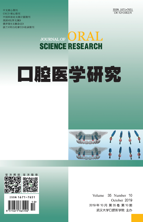
    Objective: To compare two fixed prosthesis treatment concepts supported by four implants in an atrophic maxilla. Methods: Two different treatment concept models were constructed. The conventional group was two mesial vertical implants in the lateral incisor regions and two distal tilted (45°) implants in the second premolar regions of the maxilla. The modified group was two mesial vertical implants in the lateral incisor regions and two short vertical implants in the second premolar regions of the maxilla. Numerical simulation was performed under two loading types. Results: Under the first loading type, the conventional group showed a higher stress concentration than the modified group in the bones. For the implants, the peak of von-Mises stress of the conventional group was lower than that of the modified group. Under the second loading type, the conventional group showed higher stress concentrations than the modified group, either on the bones or on the implants. Conclusion: Both treatment concepts can be applied in edentulous maxilla. Compared to the conventional group, the modified group can transmit less occlusal force to the supporting tissues.
    References | Related Articles | Metrics
    Effect of Stress State in Implant-bone Interface on Bone Osteocytes in Peri-implant Bone
    JIN Zhiheng, LI Qing
    2019, 35(10): 936-939.  DOI: 10.13701/j.cnki.kqyxyj.2019.10.007
    Abstract ( 229 )   PDF (3063KB) ( 248 )  
    Objective: To investigate the effect of stress state on bone osteocytes around dental implants using finite element analysis and animal experiment. Methods: The designed implants including full-thread and delete-thread implant were placed in proximal tibial metaphysis of rabbits. The animals were sacrificed at 7 days, 15 days, 30 days, and 60 days after surgery, and undecalcified tissue sections were prepared for SEM. The finite element models of implants and tibia metaphysis were installed and the process of implantation was simulated by computer-aided design software. The von Mises stress in peri-implant bone were recorded and analyzed. Results: The surrounding bone within 0.75 mm from surface of implant in full-thread implant group exhibited higher von Mises stress than delete-thread implant. The SEM images indicated that the number and morphological of osteocytes between two groups of implants at 7 days and 14 days were significantly different. Conclusion: The FEA can simulate the stress distribution on peri-implant bone in the initial state after implantation. The biological activity of osteocyte may be closely related to the stress state, which demonstrated a better response to stress stimuli in early stage of osseointegration and changes of number and morphology of osteocyte.
    References | Related Articles | Metrics
    Comparative Study on Effect of Treatment Methods on Dose of Subgingival Excessive Cement during Cementing the Implant Crowns.
    YU Shiqi, GAO Xu, WEN Yong, XU Xin
    2019, 35(10): 940-943.  DOI: 10.13701/j.cnki.kqyxyj.2019.10.008
    Abstract ( 275 )   PDF (1507KB) ( 277 )  
    Objective: To observe the effect of treatment methods on the subgingival excessive cement of cement-retained implant crowns. Methods: 24 die stone models of mandibular posterior denture with bone level implant were used to cement crowns by four methods, namely, cement venting hole on the occlusal (group A), on the lingual surface (group B), isolation of PTFE on subgingival region of abutments (group C), and routine cementation (group D)(n=6). To collect the subgingival residual cement, the weigh was measured by electronic balance (mg). Analysis of one-way ANOVA was conducted with SPSS 17.0 (α=0.05). Results: The weight of subgingival excessive cement in group A and group C were less than those in group B and group D (P<0.05). Neither significant difference between group A and group C nor between group B and group D was observed (P>0.05). Conclusion: Compared with the cement venting hole on the lingual side and conventional bonding method, cement venting hole on the occlusal surface and the use of PTFE membrane to isolate the surface of abutment through the subgingival area during bonding can reduce the subgingival excessive cement effectively.
    References | Related Articles | Metrics
    Effect of Modified Adhesive Retention Technique on Tooth Function in Treatment of Dentition Defect with Oral Implantation.
    LIU Zuzhi, FU Dongjie
    2019, 35(10): 944-947.  DOI: 10.13701/j.cnki.kqyxyj.2019.10.009
    Abstract ( 227 )   PDF (1039KB) ( 284 )  
    Objective: To analyze the effect of dental implant retention method on the prognosis of dentition defect. Methods: From January 2017 to January 2018, 84 patients with dentition defect treated by dental implant were divided into two groups according to double blind method. The modified plaque index, improved bleeding index, alveolar height, thickness, bone resorption, and treatment satisfaction score were compared between the two groups. Results: The alveolar height and thickness of the modified group were higher than those of routine group, and the bone resorption was lower than that of routine group (P<0.05). After 1 year follow-up, the modified plaque index of the modified group was 0, and the proportion of the first grade was significantly better than that of the routine group (P<0.05). The modified bleeding index in the modified group was better than that in the routine group (P<0.05). The scores of masticatory function, language function, firmness, comfort, and beauty in the modified group were higher than those in the routine group (P<0.05). Conclusion: Dental implant is positive for the treatment of dentition defect. The modified bonding technique is better than the conventional one in the short-term effect.
    References | Related Articles | Metrics
    Management of Nasopalatine Canal Associated with Implant Placement in Maxillary Central Incisor Sites: A Pilot Study.
    LI Zhijin, WENG Yanming, ZHOU Ronghua, DU Jiao, XIONG Caihua, DONG Qingshan, GUO Jiaping
    2019, 35(10): 948-952.  DOI: 10.13701/j.cnki.kqyxyj.2019.10.010
    Abstract ( 341 )   PDF (3894KB) ( 315 )  
    Objective: To explore the management of nasopalatine canal associated with implant placement in maxillary central sites and assess the efficacy and safety. Method: A case series retrospective study was performed between July 2016 and June 2018 of patients who were treated with implants in maxillary central incisor sites. The inclusion criteria were that cases with perforation into the nasopalatine canal during the digital treatment planning and implant osteotomy. According to the size of the perforation, different approaches were adopted to facilitate the implant placement in the compromised sites. The following parameters were assessed: neurosensory status of the anterior palate, postoperative pain and implant survival rate. Results:Of the 48 patients (71 implant sites), a total of 10 patients (10 implant sites) fulfilled the inclusion criteria. Osseointegration was achieved in all of the 10 implants. Only one patient reported minor sensory alterations in the anterior palatal zone after implant placement, which disappeared in 3 months. At the final examination (follow-up of 6-18 months), no fixtures had been lost, and no marginal bone loss exceeding 1 mm was observed at the mesial and distal aspects of any implant, and all peri-implant tissues had a healthy appearance. Conclusion: The morphology and dimensions of the nasopalatine canal should be properly evaluated prior to dental implant insertion to replace missing maxillary central incisors. If penetration into the nasopalatine duct cannot be avoided during the osteotomy following the principle of prosthetically driven implant placement in the maxillary central incisor site, proper approaches could be adopted according to the size of the penetration, and predictable effects of rehabilitation of the atrophied premaxilla could be obtained.
    References | Related Articles | Metrics
    Finite Element Analysis of Different Designs for Maxillary First Premolars with Lingual Cusp Oblique Defect.
    KANG Ting, SHI Siqiong, ZHAO Wei, ZHENG Rui, LI Xixi, XIE Jing, GUO Ling
    2019, 35(10): 953-956.  DOI: 10.13701/j.cnki.kqyxyj.2019.10.011
    Abstract ( 225 )   PDF (1505KB) ( 346 )  
    Objective: To analyze the stress distribution of the maxillary first premolar oblique defect with different methods by three-dimensional finite element analysis. Methods: Three dimensional finite element models of maxillary first premolar oblique defect were created by cone beam CT. The intact maxillary first premolar was served as the control group. Experimental groups were established according to different design. The stresses were investigated using finite element analysis software. Results: (1) The stresses of the residual dentine were increased by reduced depth of endocrowns restoration and increased elastic modulus of the material in certain range. (2) The stresses of the residual dentine in the fiber post and crown restoration of lingual canal group (C2) were smaller than that of the buccal canal group (C1) (P<0.05). The stresses of the residual dentine in the metal post and crown restoration of buccal canal group (D1) were smaller than that of the lingual canal group (D2) (P<0.05). (3) The stresses of the residual dentine of Lava Ultimate Endocrown restoration with the 4mm depth was lower than that of the lingual canal with the fiber post and crown restoration (P<0.05). Conclusion: When the maxillary first premolar is obliquely fractured, Lava Ultimate endocrown restoration with the 4mm depth may be the best restoration. If fiber post and crown restoration is choosed, lingual canal should be selected. When metal post and crown restoration is choosed, buccal canal should be selected.
    References | Related Articles | Metrics
    Clinical Effect Analysis of Glass Fiber Post Crown and Pure Titanium Post Crown
    REN Guifang
    2019, 35(10): 957-960.  DOI: 10.13701/j.cnki.kqyxyj.2019.10.012
    Abstract ( 307 )   PDF (757KB) ( 322 )  
    Objective: To analyze and compare the effect of residual crown and root's repairment by glass fiber post and pure titanium post. Methods: 235 patients (334 teeth) with residual crown or residual root in the time period of September, 2013 to March, 2017 were selected. In accordance to the admission time and patient's will, patients were divided into two teams. Patients' defect were repaired by crown and tracked by 3 years. Results: No significant difference was found on the general success rates, anterior teeth success rates, premolar success rates, and metal team's success rate (P>0.05). There was a significant difference on the anterior teeth success rate and the posterior teeth success rate of fiber team (P<0.05). On the basis of the appearance and comfort survey which responded by patients from successful cases, 94.55% patients from fiber team had feedbacks of "strongly satisfied". Such satisfaction rate was higher to the metal team. Conclusion: At current time, repairment of residual crown and root by glass fiber post and titanium post were ideal treatments.
    References | Related Articles | Metrics
    Inhibitory Effect and Acid Resistance of PDT Combined with Er:YAG Laser on Cariogenicity in Rats
    ZHANG Lei, YAO Liping, MENG Lei
    2019, 35(10): 961-965.  DOI: 10.13701/j.cnki.kqyxyj.2019.10.013
    Abstract ( 179 )   PDF (2708KB) ( 182 )  
    Objective: To investigate the bactericidal effect of photodynamic therapy (PDT) combined with Er: YAG laser on dental caries in rats and the acid resistance of dental hard tissue. Methods: 28 rats were randomly divided into 7 groups. The germicidal efficacy of each group was observed by spectrophotometer to determine the optical density (OD) value. The dissolution of calcium and phosphorus was detected after being soaked in 0.1 mmol/L lactic acid solution for 24 hours. The hardness of each group was tested by digital microhardness tester before and after lactic acid treatment. The tooth surface morphology, cleanliness, and dentinal tubules were observed by scanning electron microscope (SEM). Results: Compared with negative control group,the growth of bacteria of each group was significantly reduced. After lactic acid treatment, calcium and phosphorus were dissolved at different degrees, and the average microhardness of the surface was decreased. SEM observation showed that the surface of PDT combined with Er:YAG laser group was smooth and had obvious melting sealing effect on dentinal tubules. Conclusion: PDT combined with Er:YAG laser treatment of dental caries can not only kill cariogenic bacteria effectively,but also seal dentinal tubules significantly. It enhanced the acid resistance of tooth hard tissue and prevent the dental caries.
    References | Related Articles | Metrics
    TNF-α Increasing the Angiogenic Potential of Dental Pulp Cells and Endothelial Cells in Co-culture System.
    AN Li, SHEN Shuai, WANG Luyao, LI Yong, NIU Yumei, PAN Shuang
    2019, 35(10): 966-969.  DOI: 10.13701/j.cnki.kqyxyj.2019.10.014
    Abstract ( 214 )   PDF (2011KB) ( 246 )  
    Objective: To investigate the effects of human dental pulp stem cells (hDPSCs) and human umbilical vein endothelial cells (HUVECs) in the co-culture system on formation of endothelial tubules and vascular networks. Methods: hDPSCs were co-cultured with HUVECs at a ratio of 1∶5. Matrigel assay were performed on the co-culture system to detect the total branching lengths and the number of branches. CCK-8 assay was used to examine the effect of TNF-α on the cell proliferation. LIVE/DEAD viability/cytotoxicity kit was used to detect the mammalian cells. Results: Matrigel assay showed the total branching length. The number of branches formed in the experimental groups treated with 50 μg/L TNF-α was significantly higher than that of the control group. The decomposition time of the tubule structures was significantly prolonged (P<0.05). After treated with 50 μg/L TNF-α, there was no significant changes in the proliferation of co-cultured cells (P>0.05) . Conclusion: TNF-α enhanced the angiogenic ability of hDPSCs and HUVECs in the co-culture system in vitro.
    References | Related Articles | Metrics
    Effects of microRNA-210-3p on Proliferation and Differentiation of Human Dental Pulp Cells under Hypoxic Condition.
    YUAN Xiaoqin, HUAN Zhongping, WANG Hongtao
    2019, 35(10): 970-974.  DOI: 10.13701/j.cnki.kqyxyj.2019.10.015
    Abstract ( 185 )   PDF (2342KB) ( 208 )  
    Objective: To evaluate the effects of miR-210-3p on proliferation and differentiation of human dental pulp cells under hypoxic condition. Methods: Human dental pulp cells were isolated, and the expression of miR-210-3p was detected in human dental pulp cells under normoxia and hypoxic condition. After transfection of miR-210-3p inhibitors into human dental pulp cells, the proliferation effect under hypoxia was measured by CCK-8 assay. Alkaline phosphatase staining, real-time PCR and alizarin red staining were adopted to evaluate the regulatory effects of miR-210-3p inhibition on differentiation of human dental pulp cells under hypoxic condition. Results: Human dental pulp cells cultured under hypoxic condition were highly expressed with miR-210-3p. In addition, miR-210-3p promoted proliferation and early differentiation of human dental pulp cells under hypoxia while the late differentiation was inhibited by miR-210-3p in low oxygen. Conclusion: miR-210-3p involves in the regulation of proliferation and differentiation in human dental pulp cells under hypoxic condition.
    References | Related Articles | Metrics
    Comparison of Apically Vertical Load Induced by Three NiTi Instruments during Curved Canal Shaping in Extracted Teeth.
    XIA Zhimin, WU Changzhe, PENG Bin
    2019, 35(10): 975-978.  DOI: 10.13701/j.cnki.kqyxyj.2019.10.016
    Abstract ( 252 )   PDF (952KB) ( 283 )  
    Objective: To compare the apically vertical load induced by three NiTi instruments during canal shaping of extracted teeth. Methods: Thirty extracted mandibular premolar teeth which had single canal with a curvature between 10-20 degree were shaped with ProTape Next (PTN; Dentsply Maillefer, Switzerland), ProTaper Universal (PTU,Dentsply Maillefer, Switzerland), and M3 (China) systems (n=10 for each system). During root canal shaping, time and apically vertical load were recorded. Results: PTN group had the lowest apically vertical load, while PTU group and M3 group had no significant difference. Conclusion: ProTaper next NiTi instrument system (PTN) had the lowest values of apically vertical load while shaping, which may attributed to its safety.
    References | Related Articles | Metrics
    Expression and Significance of Beclin1 and Apoptosis Related Proteins in Oral Carcinogenesis
    SUN Weike, WANG Peiyuan, LIU Wei, WANG Xia
    2019, 35(10): 979-983.  DOI: 10.13701/j.cnki.kqyxyj.2019.10.017
    Abstract ( 218 )   PDF (5722KB) ( 238 )  
    Objective: To study the clinical significance of Beclin1 and its relationship with bcl-2, survivin, and bax in oral carcinogenesis. Methods: Immunohistochemical SP method was used to detect the expression of Beclin1, bcl-2, survivin, and bax in oral carcinogenesis tissues including oral cancer (60 cases), oral dysplasia (48 cases), and normal oral mucosa (60 cases), and to study the clinical significance of Beclin 1 in oral squamous cell carcinoma (OSCC) and its relationship with bcl-2, survivin, and bax expression in oral carcinogenesis. Results: The expression of Beclin1 was significantly increased in OSCC and normal mucosa (65% and 75%, respectively), and the positive expression rate in dysplasia significantly decreased (54.2%, P<0.05). Survivin and bcl-2 expression were higher in OSCC (73.33% and 70%) than in dysplasia and normal mucosa (P<0.05); the expression rate of bax increased in dysplasia than OSCC and normal mucosa (P<0.05). Beclin1, bcl-2, survivin, and bax expression was closely related to diameter of tumor and lymph node metastasis (P<0.05). The expression of Beclin1 was closely related to bcl-2, survivin, and bax (P<0.05; r=0.421, 0.367 and 0.542) in OSCC. Conclusion: Autophagy and apoptosis were involved in oral carcinogenesis, and were associated with invasion and metastasis of oral cancer, which synergistically participated in the development of oral cancer
    References | Related Articles | Metrics
    Effects of MicroRNA-218-5p Targeting ABCG2 Gene on Proliferation, Migration, and Invasion of Tca-8113 Cells.
    WEI Wei, TANG Yi, DAI Jing, CHEN Hujie
    2019, 35(10): 984-988.  DOI: 10.13701/j.cnki.kqyxyj.2019.10.018
    Abstract ( 208 )   PDF (1025KB) ( 204 )  
    Objective: To investigate the effects of miR-218-5p targeting ABCG2 gene on the proliferation, migration, and invasion of Tca-8113 cells and its mechanism. Methods: qRT-PCR and western blot were used to detect the expression of miR-218-5p and ABCG2 in Tca-8113 and NOK cells. The targeting relationship between miR-218-5p and ABCG2 was detected by dual luciferase reporter gene experiment. miR-218-5p mimic, ABCG2 siRNA and miR-218-5p mimic+pcDNA-ABCG2 were transfected into Tca-8113 cells by liposome method. The proliferation, migration, and invasion of Tca-8113 cells were detected by MTT and Transwell tests. Results: Compared with NOK cells, miR-218-5p was significantly down-regulated in Tca-8113 cells, and ABCG2 was significantly expressed (P<0.05). Over-expression of miR-218-5p and inhibition of ABCG2 inhibited proliferation and migration of Tca-8113 cells (P<0.05). There is a targeted binding relationship between miR-218-5p and ABCG2. Over-expression of ABCG2 reverses the inhibitory effect of up-regulated miR-218-5p on proliferation, migration, and invasion of Tca-8113 cells. Conclusion: Up-regulating the expression of miR-218-5p can target ABCG2 to inhibit the proliferation, migration, and invasion of Tca-8113 cells, which may provide a new target for the treatment of OSCC.
    References | Related Articles | Metrics
    Morphological Analysis of Dental Enamel and Root Cementum at the Cervical Region
    KONG Ninghua, SHI Shenggen, LV Yalin, LIU Lin
    2019, 35(10): 989-993.  DOI: 10.13701/j.cnki.kqyxyj.2019.10.019
    Abstract ( 228 )   PDF (3662KB) ( 267 )  
    Objective: To characterize dental enamel and root cementum at the cervical region of healthy human teeth by using field-emission scanning electron microscopy (FESEM) and atomic force microscopy (AFM). Methods: Sound human first premolars freshly extracted for orthodontic reasons were included in the study. The enamel surface on the buccal side of the tooth was analyzed in a narrow area, 1mm coronal to the zenith of cemento-enamel junction (CEJ). The cementum surface on the buccal side of the tooth was analyzed in a narrow area, 1mm apical to the zenith of CEJ. The value of Ra and Rq was measured. Results: The gross appearance of the cervical enamel was characterized by overlapping wave-like layers. The border of each enamel layer was highly undulated and formed in most areas a distinct rounded step towards the underlying layer. The exposed surface of each enamel layer was relatively smooth with very shallow depressions of Tomes' processes pits. The diameter of the Tomes' process pit was 4 to 6 microns. The cementum was characterized by projections above the general plane of the mineralizing front. The mean Ra and standard deviation in μm were: enamel, 0.459±0.223; cementum, 0.685±0.229. The mean Rq and standard deviation in μm were: enamel, 0.580±0.248; cementum, 0.796±0.261. There were statistically significant differences (P<0.05) among Ra and Rq between enamel and root cementum. Conclusion: Dental enamel surface and root cementum surface at the cervical region showed a clear difference in topography. Enamel surfaces were slightly smoother than root cementum surfaces. The findings from the study of the morphology of teeth gave us va-luable insights for designing percutaneous implants.
    References | Related Articles | Metrics
    Reconstruction of Condylar Bone and Expressions of Sox9, MMP-13, and VEGF following Rabbit Articular Disc Perforation
    XIAO Yanju, WU Guomin, ZHANG Yaoyao, CHEN Kai, WANG Xiangshen
    2019, 35(10): 994-1000.  DOI: 10.13701/j.cnki.kqyxyj.2019.10.020
    Abstract ( 190 )   PDF (8766KB) ( 89 )  
    Objective: To analyze the reconstruction of subchondral bone of TMJOA, to investigate the expression of Sox9, MMP-13, and VEGF, and to explore the mechanism of their effects on subchondral bone reconstruction. Methods: A full-thickness perforation in the diameter of 2.5 mm was made on the posterolateral side of the articular disc. BV/TV, BS/BV, Tb.Th, Tb.Sp, and Tb.N of subchondral bone were calculated by micro-CT. HE staining and immunohistochemical staining was carried out to analyze the expression and localization of Sox9, MMP-13, and VEGF, and semi-quantitative analysis was carried out. Results: Experimental group (EG) showed typical histopathological features of OA. BV/TV, Tb.Th, and Tb.N of EG were significantly decreased (P<0.05), and BS/BV and Tb.Sp of EG were significantly increased (P<0.05). The expression of Sox9, MMP-13, and VEGF in the subchondral bone of EG were significantly higher than those of sham operation group (SG) (P<0.05). Conclusion: The reconstruction of subchondral bone is an important feature of TMJOA. Sox9, MMP-13, and VEGF play an important role in bone remodeling.
    References | Related Articles | Metrics
    Cellular Compatibility and Antibacterial Properties of Magnesium Alloy after Ultrasonic Microarc Oxidation-doped Zn-HA Silane-phytic Acid Treatment.
    ZHANG Peng, ZHOU Libo, ZHANG Huiming, ZHANG Meiling, JIANG Feng, HUANG Yubo, LI Muqin, LI Dechao
    2019, 35(10): 1001-1004.  DOI: 10.13701/j.cnki.kqyxyj.2019.10.021
    Abstract ( 203 )   PDF (2587KB) ( 242 )  
    Objective: To test the cellular biocompatibility and antibacterial property of magnesium alloy after ultrasonic microarc oxidation-doped Zn-HA silane-phytic acid treatment. Methods: The magnesium alloy UMAO-silane-phytic was designed as the control group. Zn-HA at concentration of 2 g/L, 4 g/L, and 6 g/L were added respectively as the experimental group. The compatibility of different treatment membrane cells was detected by CCK-8 kit, scanning electron microscope, and laser confocal microscope. Acridine orange/ethidium bromide (AO/EB) double fluorescent staining was used to detect the antibacterial. Results: The optical density (OD) value of cell adhesion and proliferation measured by CCK-8. The OD values were in the order at: group C>group D>group B>group A. The number and shape of cells in the group C had the best extension. The antibacterial of 6 g/L Zn-HA group was obviously superior to other groups. Conclusion: The UMAO-doped 4 g/L Zn-HA silane-phytic acid treatment of magnesium alloy had the best biocompatibility. The UMAO-doped 6g/L Zn-HA silane-phytic acid treatment of magnesium alloy had the strongest antibacterial ability.
    References | Related Articles | Metrics
    Supernumerary Tooth in Left Maxillary: One Case Report
    GONG Lingwei, LIU Siling, QIU Xili, XIE Xiaoli
    2019, 35(10): 1005-1006.  DOI: 10.13701/j.cnki.kqyxyj.2019.10.022
    Abstract ( 326 )   PDF (2589KB) ( 323 )  
    Supernumerary teeth are common clinical dental anomalies, which can cause many problems like malocclusion and failure of permanent teeth eruption. Supernumerary teeth can be found in permanent dentition, or rarely in primary dentition. This article reports one case with supernumerary teeth in the left maxillary.
    References | Related Articles | Metrics